QuantCenter

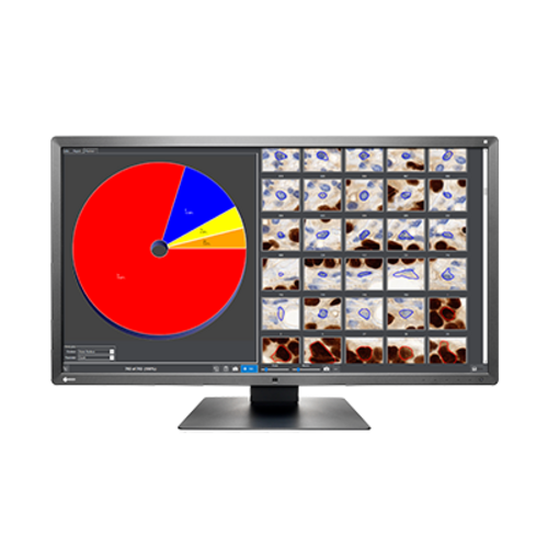
QuantCenter
- Computer-supported classification of different tissue types
- Analyse and quantify your IHC and fluorescent stainings: reproducibly and objectively
- Adapt QuantCenter to your requirements
- Benefit from the integrated report function and simple data export
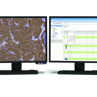
A clear benefit of digital slides is the ability to conduct computer-aided image analyses of entire tissue sections. The results calculated using software algorithms are objective, reliable and reproducible – which helps to make diagnosis easier.
QuantCenter is a repository of high-performance image analysis tools for the quantification of tissue structures, IHC staining and CISH and FISH samples.
You can call up the modules from CaseViewer and, in some cases, queue them to generate an efficient image analysis. For example, you could use the modules to analyse a specific nuclear staining in a specific tissue type on a digital slide.
The modules feature a high-performance reporting function. Alternatively, the results can be exported.
Take advantage of the opportunities of digital pathology to enable thorough and efficient analysis of your IHC or fluorescence-stained tissue sections.
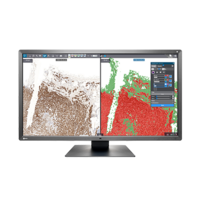
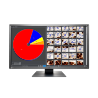
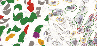
PatternQuant: Tissue segmentation and classification
- Identification, segmentation and quantification of different tissue structures
- Detailed analysis of selected tissue types with further QuantCenter modules
- Batch processing of numerous digitized slides
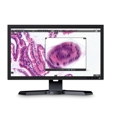
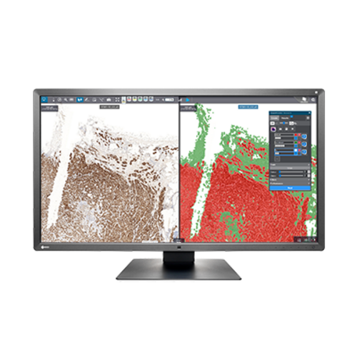
HistoQuant: Histological image analysis
- Separation of up to 10 different levels, applicable to any staining
- Measurement of morphological and densitometric characteristics

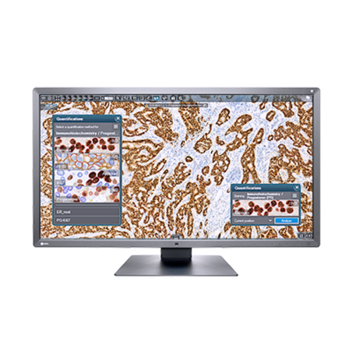
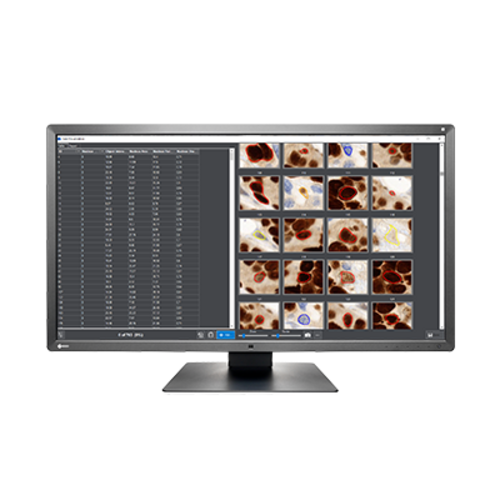
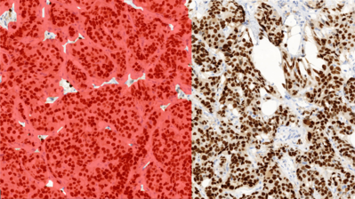
NuclearQuant: Automatic quantification of IHC nucleus staining
- Automatic evaluation e.g. of ER, PR, Ki67 and P53 staining
- Detection of cell nuclei and measurement of colour intensity in the chromogenic channel by using colour deconvolution
- Subdivision of the cell nuclei into negative to highly positive
- IVD-certified for oestrogen receptor and progesterone receptor
![]()
MembraneQuant: Automatic quantification of IHC membrane staining
- Cell membrane detected based on colour deconvolution and measurement of the chromogenic channels’ colour intensity
- Subdivision of the detected membranes into negative to highly positive
- IVD-certified for HER2 quantification

CellQuant: Immunohistochemical counting of cell nuclei, cytoplasm and membranes
- Differentiation of cell compartments though allocation to “immunostain” or “counterstain”
- Can be used for both BF and FL staining

DensitoQuant: Measurement of IHC stain intensity
- Fast and effective tool to quantify the intensity of immune staining
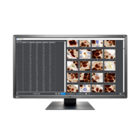
- Suitable for TMA evaluations
- Measures the stain density of an entire section in just a few minutes

CISHQuant / CISH-RNA: Analysis of chromogenic in-situ hybridisation
- Identification of CISH signals using the spot detection function (colour intensity, size, contrast)
- CISH-RNA to prove viral RNA in the infected cell nucleus
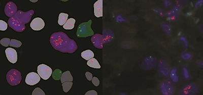
FISHQuant: Analysis of FISH samples
- Automatic or user-defined nuclei segmentation and threshold definition for FISH signals
- Specification of numerical aberrations in the stained genes, as well as classification of the nuclei into “normal”, “abnormal” and “artefact”
- Suitable for tissue samples and samples in suspension
- Intuitive determination of suitable probe types for structural aberrations, numerical deviations, locus-specific analyses or based on your own criteria
Sysmex Europe SE
Bornbarch 1
22848 Norderstedt
Germany
+49 (40) 527 26 0
+49 (40) 527 26 100