
The optional white pathological and precursor blood cell application ‘WPC’
The WPC application was designed to further classify samples initially flagged positive for ‘Blasts/Abn Lympho?’ by the DIFF application. WPC analysis excludes samples with potentially malignant cells with great specificity, which results in minimising the number of samples that have previously been flagged as potentially malignant by the DIFF analysis. The combination of DIFF and WPC analysis supports optimal differentiation between suspect malignant and suspect reactive samples.
The WPC channel can classify suspect samples into one of three clearly defined categories (‘reactive’, ‘suspected malignant’ or ‘negative’). This supports laboratories to classify all samples into one of those categories and characterise reactive conditions further, once a malignant condition has been excluded.

This refined flagging helps to streamline and accelerate the laboratory workflow as it reduces the need to perform time-consuming and expensive follow-up tests that are required when a malignant condition is suspected. Further, it supports reducing manual work (e.g., for smears) for the laboratory staff and gives them more orientation on which pathological cells to look out for in a particular sample.
The WPC application requires the presence of the WPC channel available on specific XN-Series and XR-Series analyser units and uses fluorescence flow cytometry technology.
With the optional HPC licence, the WPC channel can additionally be used to quantify haematopoietic progenitor cells.
WBC analysis with fluorescence flow cytometry
The reaction caused by the dedicated reagents in the WPC channel depends specifically on the composition of the membrane lipids. Mature white blood cells have a membrane lipid composition different to immature or reactive cells, so they are affected to a greater extent, leaving the cells in a less native stage. This makes the cell membrane more permeable for the proprietary fluorescence marker that labels the DNA of the nucleus in the next step of the reaction. The signals corresponding to cell size, internal structure and nucleic acid content are therefore directly related to the functionality of the cells.
Due to their membrane lipid composition, blasts are not permeated very strongly by the lysis reagent. Consequently, they show relatively low fluorescence signals and high forward scattered light signals because they remain mostly intact.
Neoplastic lymphocytes on the other hand are more mature and their membranes are more readily permeated, causing higher fluorescence signals and lower forward scattered light signals due to cell shrinking. These differences allow a reliable identification of such suspect malignant cells.
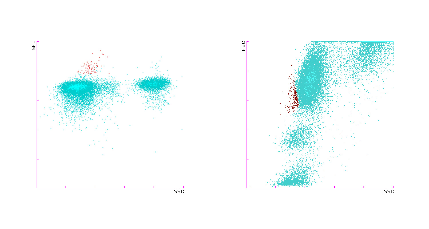
The figure below shows different views of WPC channel scattergrams. The red cluster in the scattergram on the left depicts a population of abnormal lymphocytes, while the WPC (SSC-FSC) scattergram on the right shows a blast population (red colour) present in the sample. In addition, related images of the 3D scattergrams (XR-Series only) are shown below.
WPC Scattergram
WPC Blast Scattergram
Benefits for laboratories and clinicians
- WPC analysis helps to minimise the number of samples that are finally flagged as potentially malignant and so simplifies the laboratory workflow by reducing the need for time-consuming and costly tests required when a malignant condition is suspected.
- You can have confidence in performing smears on the right samples without losing time spent on false-positive ones. Smear review is accelerated by focussing on specific cell types.
- WPC analysis is performed as an automated reflex test, which provides you with increased walk-away time.
- Measurement of reactive cells (Extended Inflammation Parameters, optional licence) allows clinicians deeper insight into the immune response status, particularly after having excluded malignant conditions specifically with WPC analysis.
- With the optional HPC licence, you can quantify haematopoietic progenitor cells, which can add to the lab service that supports haematopoietic stem cell transplantations (HSCT) on site with determining the optimal time point for apheresis procedures.
Documents for download
-
Lab card: Further classification of WBC abnormalitiesPDF (542 KB)
-
Lab card: Supporting the apheresis ward with excellent lab servicePDF (216 KB)
-
Lab card: From cell count to cell functionalityPDF (666 KB)
-
White paper: Sensitive assessment of WBC functionality and greater workflow efficiencyPDF (2 MB)
-
White paper: Going beyond the visiblePDF (2 MB)


