The optional reticulocyte count application ‘RET’
The RET application on Sysmex’s five-part differential haematology analysers uses a dedicated analysis channel for the quantification of reticulocytes (RET; absolute count and percentage), and also quantifies mature and immature reticulocytes with ease. Besides the RET count, it delivers the immature reticulocyte fraction (IRF) plus a remarkable range of further parameters that have shown* considerable value in supporting diagnoses of red blood cell disorders. In particular, the RET application delivers valuable support in anaemia diagnostics. The appropriate combination of parameters highlights the complete picture of erythropoiesis and its further development.
RET-He is one of the advanced clinical parameters that is derived from the RET analysis. It quantifies the haemoglobinisation of the reticulocytes and has proven of being superior to all mature red blood cell-related parameters*.
Further parameters of interest from the RET application cover hypo- and hyperchromic red blood cells (HYPO-He / HYPER-He), the difference in the haemoglobinisation of reticulocytes versus that of all red blood cells (Delta-He), the reticulocyte production index (RPI)** and fragmented red cells (FRC)**.
If you want to go beyond blood parameters and streamline your haematology sample workflow, then team up the RET application with the Extended IPU:
There may be various reasons behind an increased MCHC, some of which are related to true RBC disorders, whereas many others stem from measurement interferences. The optional CBC-O** add-on based on the research of Berda-Haddad et al. embedded in the Extended IPU can support you effectively. It delivers conclusive information about the cause of interference, suggests replacement of the affected parameters with their counterparts from the RET analysis (e.g., RBC-He, RBC-O**, HGB-O**), and automatically recalculates the RBC indices. Using the RET channel together with CBC-O reduces the need for laborious manual procedures of such samples.
The RBC Defect Workflow Optimisation (RWO)** rule set (optional) in the Extended IPU combines the CBC-O rule set, the RBC score and advanced RBC and RET parameters. The intelligent two-step algorithm, which is based on the research of Nivaggioni et al.* and Berda-Haddad et al.*, aims to support the identification of patients with hereditary RBC diseases and iron deficiency anaemia (IDA). It achieves an improved laboratory workflow by forwarding samples suspected of hereditary RBC diseases to the respective tests that can confirm the diagnosis and at the same time correctly exclude IDA samples that can generate spurious results.
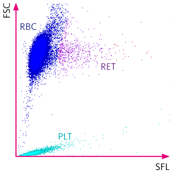
Blood cell analysis including reticulocytes with fluorescence flow cytometry
In the RET channel, a specific reagent slightly perforates the cell membranes of red blood cells and platelets, and so allows the fluorescence marker to penetrate the cells while leaving them largely intact. The fluorescence marker labels nucleic acids and other components of reticulocytes and platelets, whereby the intensity of the resulting fluorescence signal is directly proportional to the nucleic acid content.
Reticulocytes emit a higher fluorescence signal than mature red blood cells, which no longer contain RNA. Using primarily the fluorescence signal for differentiation, together with the forward scattered light the reticulocytes can be separated from mature red blood cells and platelets and are displayed accordingly in the RET scattergram.
According to their fluorescence intensity, reticulocytes are fractionated into three categories:
- LFR (low-fluorescence ratio)
- MFR (medium-fluorescence ratio)
- HFR (high-fluorescence ratio)
The IRF (immature reticulocyte fraction) reflects the proportion of immature reticulocytes and is calculated from the sum of MFR plus HFR.
The RET application also provides the optical platelet count (PLT-O) as platelets also contain intracellular RNA. Automated reflex measurement is implemented for samples with unreliable impedance platelet (PLT-I) counts since the use of fluorescence flow cytometry resolves many PLT-I interferences that are based on volume overlaps.
For more information on how the advanced clinical parameters mentioned earlier are derived and determined, and how they can be put to use, visit their dedicated ‘Sysmex parameters’ pages.
The below figure shows the scattergram of the RET channel with a normal cell distribution.
Benefits for laboratories and clinicians
- Rapid determination of the erythropoietic status using haematological routine diagnostics.
- The combined information of quantity- and quality-related RET parameters provides a comprehensive picture of erythropoiesis and its further development.
- The RET profile can be analysed in whole blood mode (standard) as well as in the pre-diluted mode. This helps with samples containing very low blood volumes.
- An optical platelet count (PLT-O) is part of the reticulocyte analysis using fluorescence flow cytometry, which resolves many interferences affecting the impedance platelet count (PLT-I). It is therefore implemented as an automated reflex analysis for samples with unreliable PLT-I counts.
- The optional CBC-O** add-on of the Extended IPU greatly improves the workflow for samples with an increased MCHC as it reduces manual procedures for finding the root cause of the abnormality.
- The optional RWO** add-on of the Extended IPU offers an opportunity to streamline the laboratory workflow and support in choosing the appropriate confirmatory tests for suspicious RBC disease samples.
- When compared with the haemoglobin value or other RBC parameters, the RET-He value and advanced parameter combinations enable significantly faster insight into changes in the erythropoietic status, supporting clinicians in controlling iron and/or erythropoietin therapy.
- Supports the possibility of close therapy monitoring, which is necessary, for example, with dialysis patients, or helps clinicians to rapidly identify an emerging anaemia as a side-effect or even the recovery during successful treatment.
- The IRF (immature reticulocyte fraction) is an indicator of erythropoiesis and correlates well with the engraftment of neutrophils as published* by researchers. It has also been incorporated into algorithms for the screening of HS patients*.
* For references to independent publications, please visit our publications or contact your local Sysmex representative.
For Sysmex white papers, please visit our Scientific documents library.
** Research parameter / For research use only
Documents for download
-
Clinical information card: Direct assessment of the availability of iron for haemoglobinisationPDF (300 KB)
-
Clinical information card: Information on iron deficiency early onPDF (319 KB)
-
Lab card: Valuable support in anaemia diagnosisPDF (660 KB)
-
Lab card: Get the best platelet result for each sample without delayPDF (2 MB)
-
Lab card: Your hereditary RBC defect workflow streamlined – based on the latest researchPDF (936 KB)