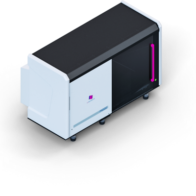
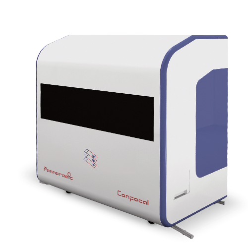
Pannoramic Confocal
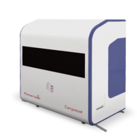
Pannoramic Confocal – 3D images in excellent quality
- Confocal scanning of the entire specimen
- High light output with minimal specimen bleaching
- Detailed view of cell nucleus thanks to 3D display
- 100 µm section thickness
- Bright-field and fluorescent scans in a single device
With the Pannoramic Confocal, 3DHISTECH presents the first – and only – confocal whole-slide scanner for pathological research.
The confocal scanning technology sequentially illuminates the entire specimen, thereby generating exceptionally high-quality images of your tissue sections. Specimens with a thickness of up to 100 µm can be scanned with the Pannoramic Confocal and then examined in three dimensions using the 3D-view software. This provides you with a particularly comprehensive view of the tissue area to be analysed.
To fulfil as many varied requirements as possible, the scanner can both generate 3D visualisations of fluorescence specimens and perform bright-field scans. With a capacity of 12 slides and an optional automatic scanning function, the Pannoramic Confocal integrates perfectly into a research laboratory.
Software
Pannoramic control software – included
The Pannoramic control software is the user interface to control your Pannoramic slide scanner. This software supports your daily work processes and the handling of the Pannoramic Confocal.
To optimise the workflow, all scanners automatically detect the specimen, focus on the tissue and create a preview on the screen. A photograph of the slide label is also created or its barcode is read. This supports archiving, saves time and reduces costs.
- Easy to use
- Choice of manual or automatic scan mode
- Automatic tissue detection
- Automatic focus
- Photograph of the slide label for documentation
- Optional barcode detection
CaseViewer – included
The CaseViewer is pre-installed on the computer linked to the slide scanner. This software not only allows the digitised tissue specimen to be viewed, but also offers a wide range of processing options. Markings can be created, comments added and several tissue specimens can be viewed in parallel in different colours, to name but a few of the features. To put it simply, the CaseViewer is your entrance to the world of virtual pathology.
| Technology | Bright-field/7-channel fluorescence 16-bit cooled monochrome sCMOS camera Objectives: 20x or 40x (optional) Maximum resolution: 31x or 62x (depending on objective) Barcode scan: 1D and 2D Bright-field and fluorescence objective: Solid-state light engine, 15,000-hour service life |
| Capacities | Specimen capacity: 12 Accepted specimen formats: 25 x 75 mm, 1 mm thickness |
| Digital storage | 3DHISTECH format (MRSX), can be coded for JPG, JPG2000 |
| Dimensions (W x D x H) | Pannoramic Confocal: approx. 95 cm x 57 cm x 100 cm |
| Weight | Approximately 50 kg |
Sysmex Europe SE
Deelböge 19 D
22297 Hamburg
Germany
+49 (40) 527 26 0
+49 (40) 527 26 100