
Bladder cancer diagnostics and surveillance
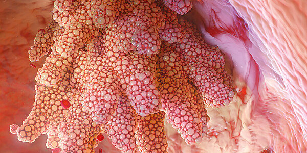
Urothelial carcinoma, also known as transitional cell carcinoma (TCC), is the most common type of bladder cancer. While it first develops in the urothelial cells that line the inside of the bladder, the initial diagnosis often only occurs once related symptoms that are characteristic of later stages of the tumour growth appear. This negatively affects the chance of successful treatment and requires invasive procedures, such as partial or complete resection of the bladder.
Urothelial carcinomas show a high rate of remission, requiring strict surveillance schemes of treated patients including multiple invasive cystoscopies.
The detection of urinary atypical cells (Atyp.C) by urinary flow cytometry bears the potential to support cancer surveillance, aiming to reduce unnecessary invasive cystoscopies.
Urinary atypical cells
Atypical cells with changes or cells that are suspected of malignancy are released from urothelial tissues through exfoliation and present with certain morphological features, which can include:
- Enlarged nucleus > 25 µm
- Increased N:C (nucleocytoplasmic) ratio > 80 %
- Irregular shape
- Hyperchromasia
- Irregular chromatin patterns
- Nucleolic hypertrophy > 5 µm
- Increased number of nucleoli
- Irregular nuclear shape
- Appearance of cellular masses

How to detect atypical cells in urine?
Urinary flow cytometry supports the detection of atypical cells by using specific fluorochromes for the staining of surface structures and nucleic acids. Marked cells are separated, detected and identified by hydrodynamic focussing, laser scanning and scattergram fingerprinting. Atypical cells are reported as research use only (RUO) parameter Atyp.C.
A potential workflow for Atyp.C
Atyp.C might help to distinguish between urine specimens with and without signs of cellular atypia using dedicated cut-off values. If samples are negative, this may preclude the need for further diagnostics, whereas positive samples will require specific follow-up diagnostics.

Bladder cancer diagnostics in research
Shukuya et al. (2023) investigated the diagnostic performance of the Atyp.C parameter on the UF-Series, concluding that it is a useful parameter in routine urine testing.
Karaburun et al. (2023) demonstrated the potential of the Atyp.C parameter in the surveillance of NMIBC patients in a prospective study. Significantly different Atyp.C values have been observed between malignant and non-malignant individuals and between those with high- and low-grade recurrence.
Aydin et al. (2021) presented a case of high-grade urothelial carcinoma detected by cellular atypia, applying the Atyp.C parameter from urinary flow cytometry in combination with automated microscopy among the study population in an outpatient setting.
Tınay et al. (2020) evaluated the Atyp.C parameter in supporting bladder cancer diagnostics in a retrospective manner in a heterogenous study population. An acceptable sensitivity of 75% and a specificity of 100% demonstrated potential value for diagnostic decisions in follow-up cystoscopy for patients with low-risk non-muscle invasive bladder cancer (NMIBC) with a potential impact on subsequent diagnostic decisions, such as the need for invasive cystoscopies.
Ren et al. (2020) demonstrated the predictive power of Atyp.C for patients with a suspected diagnosis of urothelial carcinoma. The Atyp.C findings were in agreement with cytopathology in 73% of the investigated cases, implying a potential utility of the Atyp.C parameter as an accessory test for urothelial carcinomas in the context of routine urinary diagnostics.

Scientific references
[1] Shukuya, K., Morita, Y., Hisasue, T. et al. (2023): Comparison of the clinical performance of the Atyp.C parameter of the UF-5000 fully automated urine particle analyzer with that of microscopic urine sediment analysis. Practical Laboratory Medicine 36:e00328.
[2] Karaburun, M.C., Özkaya, M.F., Ergüder, B.İ. and Süer, E. (2023): ): Investigation of Atypical Cell Parameter in the Surveillance of Patients with NMIBC; Initial Outcomes of a Single Center Prospective Study. J Med Syst 47(1):41.
[3] Aydin, O. (2021): Atypical cells parameter in Sysmex UN automated urine analyzer: feedback from the field. Diagn Pathol Diagn Pathol
[4] Tınay İ , Şahin B, Saraçoğlu S et al. (2019): “Atypical Cell’’ Parameter in Automated Urine Analysis for the Diagnosis of Bladder Cancer: A Retrospective Pilot Study. Bull Urooncol
[5] Ren, C., Wang, X., Yang, C. et al. (2020): Investigation of Atyp.C using UF-5000 flow cytometer in patients with a suspected diagnosis of urothelial carcinoma: a single-center study. Diagn Pathol
[6] Sanli, O., Dobruch, J., Knowles, M. et al. (2017): Bladder cancer. Nat Rev Dis Primers